By Paul T. Finger, MD
Description
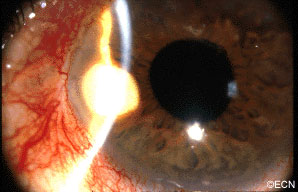
A rare complication of cataract surgery, the “pearl-cyst” may grow from on and within the iris. The tumors are thought to be caused by displaced conjunctival or epidermal epithelium. Rupture of these mucous-containing cysts (within the eye) can cause severe glaucoma.
Symptoms
Most patients present due to ocular inflammation. They may notice the white tumor growing
on the iris.
Diagnosis
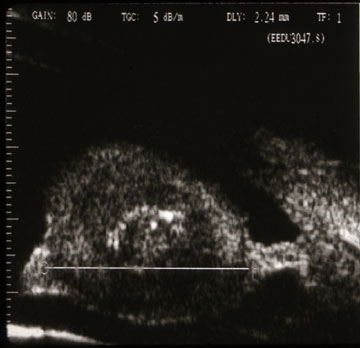
The diagnosis of “Pearl Cyst” should be considered when a patient who has had intraocular surgery (cataract, corneal transplant, or filtering for glaucoma) presents with a slowly
enlarging white iris stromal tumor. High-frequency ultrasound imaging will show three layers: an outer mantle, a low reflective mucus
layer and a central, high-reflective cholesterol crystal core.
Treatments
When “Pearl-cyst” of the iris is suspected, complete surgical excision is warranted. Though difficult, the surgeon should try to remove the cyst intact. Should the cyst rupture during removal, the surgeon should be ready to aspirate or removal the cyst contents. The mucoid contents have been reported to cause secondary glaucoma. If the outer epithelial layer, the mantle, is left behind one runs the risk of cyst recurrence.


